Understanding the source of physical discomfort is an important step in developing an effective and personalized care plan. While symptoms such as stiffness, tension, or limited mobility can provide clues, they do not always reveal the full picture. Imaging technology plays a key role in helping providers look beneath the surface and gain a clearer understanding of what may be contributing to those symptoms.
This article explains how imaging technology is used, what types of imaging may be involved, and what you can expect during the process.
Why Imaging Technology Is Used in Evaluations
Imaging allows providers to view internal structures such as bones, joints, muscles, and soft tissues. This can help identify patterns or irregularities that may not be visible during a physical examination alone.
Using imaging as part of an evaluation can help:
- Provide a clearer view of internal structures
- Support more accurate assessments
- Guide decision making for next steps
- Reduce uncertainty when symptoms are unclear
Rather than relying on guesswork, imaging provides additional data that can support a more informed approach.
Common Types of Imaging Technology
Different imaging tools are used depending on the area being evaluated and the type of information needed. Each method offers a unique perspective.
MRI (Magnetic Resonance Imaging)
MRI is commonly used to evaluate soft tissues such as muscles, ligaments, and discs.
It may be helpful for:
- Viewing soft tissue structures
- Identifying areas of inflammation or irritation
- Evaluating joints and spinal structures
MRI does not use radiation and provides detailed images of internal tissues.
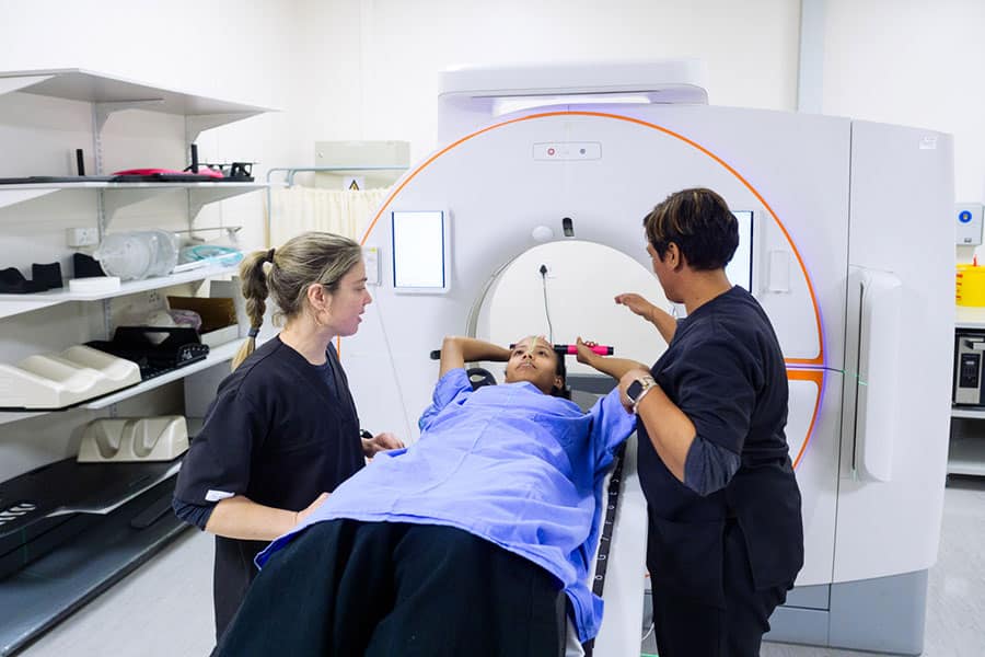
CT Scan (Computed Tomography)
CT scans use a series of X-ray images to create detailed cross-sectional views of the body.
They are often used for:
- Examining bone structures
- Identifying structural changes
- Providing detailed images of specific areas
CT scans can be especially useful when a more detailed view of bone anatomy is needed.
X-Rays
X-rays are one of the most commonly used imaging tools and are often part of an initial evaluation.
They are typically used to:
- Assess bone alignment
- Identify structural changes
- Provide a quick overview of an area
While X-rays do not show soft tissue in detail, they can provide important baseline information.
Ultrasound Imaging
Ultrasound uses sound waves to create real-time images of soft tissues and movement.
It may be used for:
- Observing muscles and tendons in motion
- Guiding certain procedures
- Evaluating specific soft tissue areas
Ultrasound is often used in dynamic assessments where movement is important.
How Imaging Helps Identify the Source of Discomfort
Imaging technology supports the evaluation process by offering visual insight into internal structures. This can help providers connect symptoms with potential underlying causes.
Correlating Symptoms with Structural Findings
Symptoms such as localized discomfort or limited movement can sometimes be linked to specific structural patterns seen on imaging. This helps create a more complete understanding of the situation.
Supporting Clinical Assessments
Imaging is typically used alongside a physical evaluation and medical history. Together, these elements provide a more comprehensive view than any single method alone.
Guiding Next Steps
Once imaging results are reviewed, providers can use that information to guide recommendations. This may include additional evaluation, changes in activity, or other next steps based on the findings.
What to Expect During an Imaging Appointment
The process varies depending on the type of imaging being performed, but most appointments follow a similar structure.
Preparation
In many cases, little preparation is required. You may be asked to:
- Remove metal objects for certain scans
- Wear comfortable clothing
- Provide a brief medical history
Your provider will give specific instructions based on the type of imaging scheduled.
During the Procedure
Each imaging method has a different experience:
- MRI involves lying still inside a machine for a period of time
- CT scans are typically quick and involve minimal movement
- X-rays are fast and completed in a few minutes
- Ultrasound is performed using a handheld device over the skin
Most imaging procedures are non-invasive and completed on an outpatient basis.
After the Appointment
Once imaging is complete, the results are reviewed and interpreted. Your provider will typically discuss findings with you and explain how they relate to your symptoms.
Limitations of Imaging Technology
While imaging is a valuable tool, it is only one part of the evaluation process.
Important considerations include:
- Not all findings directly correlate with symptoms
- Some structural changes may be common and not cause discomfort
- Imaging results should always be interpreted in context with your history and physical assessment
This is why imaging is used as part of a broader evaluation rather than as a standalone solution.
When Imaging May Be Recommended
Imaging may be considered in situations such as:
- Ongoing discomfort without a clear cause
- Changes in symptoms over time
- Limited improvement with initial approaches
- Need for a more detailed assessment
Your provider will determine if imaging is appropriate based on your specific situation.
How Imaging Supports a Personalized Approach
By combining imaging results with a detailed evaluation, providers can develop a plan that is tailored to your needs. This approach helps ensure that recommendations are based on both clinical assessment and visual data.
This process supports:
- More targeted decision making
- Better understanding of contributing factors
- Clear communication about next steps
Imaging technology plays an important role in helping identify the source of discomfort by providing a closer look at internal structures. When used alongside a thorough evaluation, it can offer valuable insight that supports informed decision making.
Understanding how imaging works and what to expect can help you feel more prepared and confident as you explore your options.